
搜集國內(nèi)外高校在線教學(xué)資源
Cancer is a scary word, but as you have learned by now, words give you the information you need to make knowledgeable decisions in consultation with your family physician and oncologist (cancer specialist).
Many cancer terms are unique to the field of oncology (study of tumors) and don’t lend themselves easily to the prefix, root, suffix system used in the previous modules. Instead, terms will be grouped and defined in broad categories such as tumor types, causes and treatments. In place of a quiz there will be a simulated case that reinforces frequently used terms.
| Good news | Bad news |
|---|---|
| Benign | Malignant |
| Low grade | High grade |
| Radiosensitive | Radioresistant |
| No metastases | metastases |
| Well differentiated | Poorly differentiated |
| Negative nodes | Positive nodes |
| In remission | Relapse |
| Surgically resectable | Inoperable |
Malignant vs. benign (literally, “evil” versus “good”)
Tumors are masses of cells that have slipped the bonds of control of cell multiplication. Malignant tumors, cancers, are life-threatening because they are invasive (spread into surrounding organs) and metastasize (travel to other areas of the body to form new tumors). Specifically, invasiveness results in penetration, compression and destruction of surrounding tissue causing such problems as loss of organ function (liver, kidneys), difficulty breathing (lungs), obstruction (intestines), possible catastrophic bleeding and severe pain.
Carcinoma is the most common form of cancer. Carcinoma develops from sheets of cells that cover a surface (example: skin) or line a body cavity (example: glandular lining of stomach). Some names for tumors of this type would be: adenocarcinoma of the prostate, adenocarcinoma of the lung, gastric adenocarcinoma, hepatocellular carcinoma (what organ is involved?). Note that the term carcinoma typically appears in the name.
A rare form of cancer arises from connective and supportive tissues, examples: bone, fat, muscle, and other connective tissues. Some names of this type of tumor would be: osteosarcoma (malignancy of bone), liposarcoma (fat) and gastrointestinal stromal tumor. Note that the term sarcoma does not always appear in the name.
 Tumor biopsies (tissue samples) are examined microscopically to determine the type and degree of development. A grading scale is used, usually Grade I to Grade IV, to describe tissue differentiation. Tumors that are well differentiated (it still looks like the original source tissue) generally have a good clinical outcome. Tumors that are poorly differentiated (the tissue has taken on a more primitive structure and may not resemble its original tissue) generally have a poorer outcome. The clinical stage of a tumor is determined by physical exam (Can you feel the tumor? Can you palpate (feel) lymph nodes? Is the tumor fixed in place (adherent to other structures)? Imaging (CT, MRI) is also an essential tool. The stage of the tumor determines if the tumor has invaded surrounding tissue, involved lymphatics (drainage channels for cell fluids other than blood) and whether the cancer has metastasized to other sites in the body.
A staging system using the letters T, N, M is also used in conjunction with Grading. “T” indicates size of tumor; “N” whether the cancer has spread into lymph nodes; “M” whether cancer cells have metastasized to other organs and areas. For example, a melanoma T2N0M0 describes a skin cancer that is between 1.0 and 2.0 mm in thickness, but has not spread into lymph nodes or other areas of the body.
Grading and staging tumors are important ways to predict the “prognosis” (progress and outcome of the disease), and which types of treatments may most likely succeed. In general, low grade tumors that have not invaded tissues, have not involved lymph nodes (negative nodes) and have not metastasized would be expected to have a better prognosis than a high grade tumor that has invaded tissues, has invaded lymphatics (positive nodes) and has metastasized. However, the prognosis of any individual patient is much more complicated than described here. Complicating factors include the general health of the patient, the effectiveness of their immune system and available treatment options. Some tumor types are very “aggressive” and are highly resistant to treatment.
Any injury to DNA (the genetic code) may result in the loss of cell cycle control, leading to uninhibited cell division. Carcinogens are cancer causing agents. Broad categories include radiation, chemicals, drugs and viruses. Don’t panic! Your once a year dental X-ray and common cold and flu viruses will not cause cancer. However, excessive radiation from nuclear to sunlight can significantly increase your risk of malignancy. The Human Papilloma Virus (HPV) is the major cause of cervical cancer. Environmental chemicals found in tobacco smoke, automotive exhaust, toxic emissions from factory smokestacks and asbestos exposure are all carcinogenic.
Curious about your risk for common cancers? Check in at Your Disease Risk at Washington University School of Medicine.
Tumor markers are substances that are produced by tumors or the body’s response to presence of a tumor. Tumor markers found in various body fluids, such as the blood, can be useful in the detection and response to treatment of certain cancers. However, most tumor markers are not specific for cancer and they may be present or even elevated with benign diseases. The absence of a tumor marker can also be useful in confirming successful cancer treatment; whereas an increase in the tumor marker level may indicate recurrence. Two well known markers are Prostate Specific Antigen (PSA) for prostate cancer and CA-125 for ovarian cancer.
It is ironic that the same agent that can cause cancer can be used to destroy cancer, but a common mechanism is at work. Fairly low to moderate doses of radiation can cause DNA damage, which may result in the malignant transformation of normal cells into cancer cells. But, high dose radiation focused on cells can destroy the cancerous cells. However, even with highly focused radiation treatment, normal surrounding tissues are exposed to the radiation and may lead to secondary cancers.
Some terms you will hear about are:
Radiosensitive – cancer degenerates in response to radiation
Radioresistant – the cancer may have a partial response or doesn’t respond at all
Fractionation – a treatment radiation dose is broken down into multiple exposures over several weeks to minimize side effects
Perhaps nothing short of surgery strikes fear into our hearts more than being told, “You’re going to need chemo”. Stories of hair falling out and nausea and/or diarrhea are awful. But, the essential action of most chemotherapeutic agents is to kill or stop the development of rapidly dividing cells. However, chemotherapy works systemically (affects the whole body) so any rapidly dividing cell, cancer or not, is affected by the medication; such as hair follicles and the lining cells of our stomach/intestines. Make sense?
Another side effect of chemotherapy is myelosuppression, where the rapidly dividing bone marrow cells are killed off. Patients may complain of extreme fatigue due to anemia (reduced number of erythrocytes) and can be at increased risk of infectious disease (reduced number of leucocytes).
 Chemotherapeutic agents that you will likely hear about are: Cisplatin, Carboplatin, Bleomycin, 5-fluorouracil, methotrexate, Vincristine, Vinblastine, and Taxol. Since the same mechanism that kills a malignant cell or blocks development of a malignant cell can have similar effects on a normal, rapidly dividing cell, any of these agents can have unpleasant side effects. Some forms of cancer treated with chemotherapy may cause the cancer to “disappear” for awhile although not cured and the patient may be symptom free sometimes for months or years. This period of holding the cancer in check is called a “remission”. Unfortunately, many such cancers, such as leukemia, reoccur and the patient is said to have “relapsed”.
Every year, promising new treatments are being developed. One of the newest is an angiogenesis (blood vessel growing) inhibitor. Medications such as Avastin and Sutent block blood vessels from growing into a tumor thereby starving the growth.
In my opinion, the best way to get rid of a cancer is cut it out. I want rid of it now! However, some tumors are so enmeshed in normal tissues that they cannot be safely cut out without severe damage to normal tissues, in other words, they are “inoperable”. And, depending upon the location (brain, prostate, etc) and the amount of excised tissue, one may be left with severe disability. However, surgery can be a complete cure for some types of tumors if done early, such as malignant melanoma (skin cancer). The probability of a cure may be enhanced after surgery by following up with additional treatments such as chemotherapy, radiation therapy or both. The term for this is “adjuvant therapy”.
Some surgical terms you will hear:
Cryosurgery – destroying malignant tissue by freezing it with a cold probe. Often used for soft tissues like liver or kidney.
Fulguration – “Lightning” in Latin. Malignant tissue is destroyed with an electrocautery instrument (electric current).
Excisional biopsy – simultaneous tissue sampling and removal of a tumor with a safe margin of normal tissue. Frequently done with suspicious skin lesions; example, malignant melanoma.
Resect- to cut and remove a segment of an organ containing a tumor.
En bloc resection – removal of the tumor and any surrounding organs or tissues that may be involved. This is often necessary for large abdominal sarcomas.
Unfortunately, not all cancer treatments are curative. Palliative treatment gives relief of symptoms, but does not cure and is reserved for advanced malignancy.
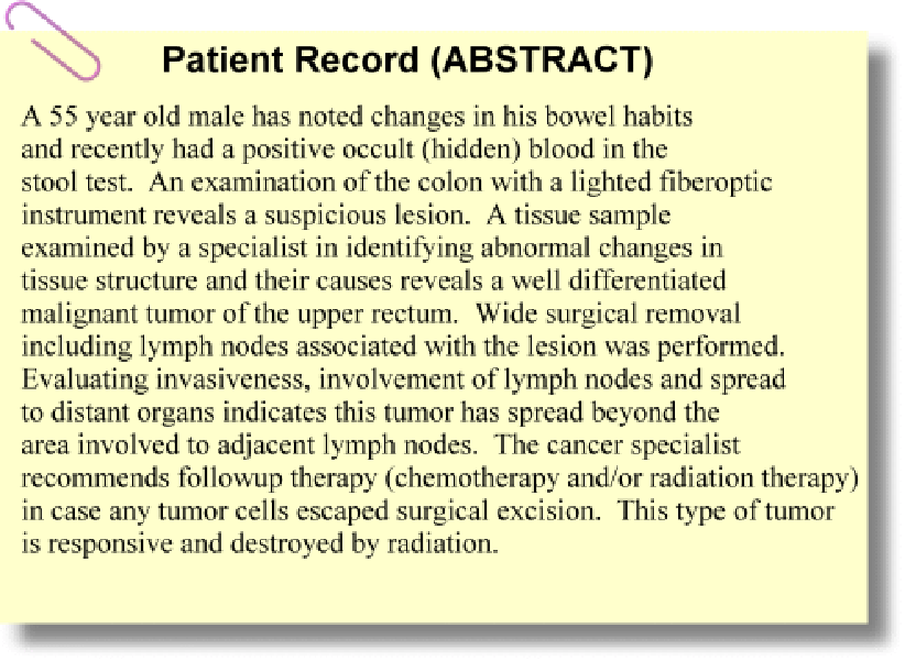
The following is a simulated patient case.
Think to yourself what each italicized term means and how it may affect the patient’s prognosis. You may find a couple of terms from previous modules. Remember, this is a cumulative learning experience!

| Good news | Bad news |
|---|---|
| Benign | Malignant |
| Low grade | High grade |
| Radiosensitive | Radioresistant |
| No metastases | metastases |
| Well differentiated | Poorly differentiated |
| Negative nodes | Positive nodes |
| In remission | Relapse |
| Surgically resectable | Inoperable |
I am optimistic! How about you? Of course, physicians don’t use the simple-minded scorecard shown above. It is just my mechanism to summarize significant information that can affect prognosis.
